


Embryo selection is a decisive phase in a fertility treatment. In fact, the success of the treatment will largely depend on it.
In the past ten years, assisted reproduction techniques have made significant advances. Thanks to this, each phase of the process has improved: from obtaining samples to embryo transfer and the subsequent implantation of the embryo, leading to the birth of a healthy baby.

Embryo selection based on morphology and kinetics has been enhanced by the development of time-lapse technology, available in our clinic's laboratory.
Monitoring embryo development with time-lapse technology allows visualisation of their evolution without altering incubation conditions. This, combined with the evaluation by the embryology team, helps determine which embryos have the highest chance of implanting and achieving pregnancy.
Selecting embryos for transfer using objective criteria has significantly improved treatment success rates, as it enhances both implantation rates and the number of healthy babies born.
In our laboratory, we use GERI and Embryoscope time-lapse incubators.
GERI is an incubator with a time-lapse system that monitors embryos from the oocyte stage to the blastocyst stage (day 5/6 of development).
The Embryoscope® time-lapse system includes a camera and image capture system that allows real-time observation of embryo development.
GERI is an incubator with a time-lapse system that monitors embryos from the oocyte stage to the blastocyst stage (day 5/6 of development).
The Embryoscope® time-lapse system includes a camera and image capture system that allows real-time observation of embryo development.
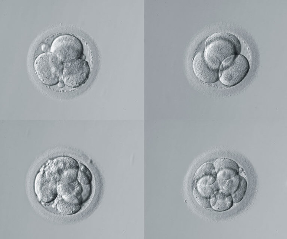
The embryology team is responsible for selecting the best embryo. They monitor the embryonic development daily. The embryos selected for transfer will be those with the highest chances of implanting. To select the best embryo, embryologists follow several criteria:
Thanks to technological advances, the tools available to embryologists for proper embryo selection have significantly improved. This allows for higher implantation rates and healthier baby births.
At IVF-Life, we use the most advanced preimplantation genetic analysis techniques for embryo selection based on genetic criteria.
We have a genetic diagnosis program designed to precisely detect possible alterations in the 23 pairs of chromosomes that make up each embryo cell. By selecting embryos without chromosomal abnormalities, the birth rate of healthy babies increases. Most importantly, by reducing the rate of miscarriages caused by genetic factors.
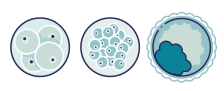
Through embryo biopsy, a few cells are extracted from the developing embryo (around day 5 of growth). Genetic material is obtained from these cells to analyse their chromosomal status. Around day 5, embryo cells have separated to form two distinct cell masses: one is the embryoblast (inner cell mass), which will develop into the future baby, and the other is the trophectoderm, which will form the placenta.
In our fertility clinics, we perform a trophectoderm biopsy, minimising risk to the embryo, as these cells are quickly replaced. In most European clinics, cell extraction for analysis is usually done on day 3 of embryonic development. This can negatively affect embryo viability, as the number of cells is still very low and not yet differentiated, leading to less reliable diagnoses.

Find answers to your questions

At IVF-Life, we have state-of-the-art technology that allows us to maintain high pregnancy rates by transferring the highest-quality embryo.
Both the medical and embryology teams do not recommend multiple embryo transfers. A twin pregnancy can lead to complications for both mother and baby, such as premature birth or intrauterine growth restriction.
Thanks to the strategies and techniques such as embryo selection, we have specialised in managing the most complex cases.


