By Vaia Siapka (Gynaecologist – Reg. No. 030314546).
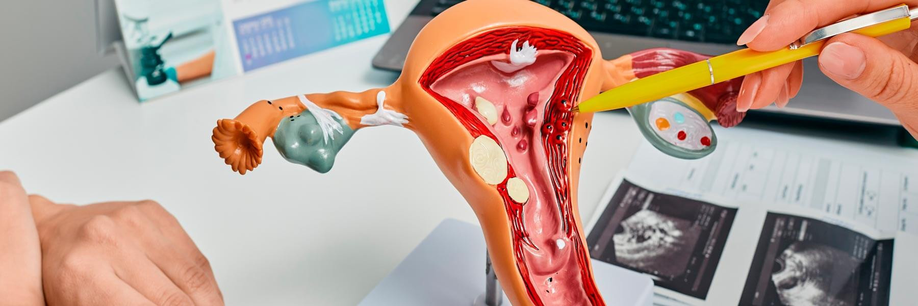
Patients undergoing egg donation treatment have an important physical advantage: the process is generally much easier than in vitro fertilisation (IVF) using their own eggs.
This is because they do not need to undergo ovarian stimulation or egg retrieval, as both procedures are carried out by the donor. Rather than stimulating her ovaries or undergoing egg collection, the patient receiving the embryo only needs to prepare her endometrium so that it is in the best possible condition at the time of transfer.
This is known as endometrial preparation for egg donation. It involves administering a combination of oestrogen and progesterone, alongside ultrasound monitoring to assess how the endometrium is responding. The treatment therefore involves fewer procedures and offers greater scheduling flexibility, although the protocol and timings must always be tailored to each patient.
Before embryo transfer, the endometrium must be prepared to ensure that the lining of the womb is in a suitable condition and, crucially, that the timing of transfer is synchronised with the embryo's stage of development.
There is no single protocol that suits every woman. For patients who still ovulate, preparation may be based on their natural menstrual cycle. In other cases, a hormone replacement or programmed cycle is used, with oestrogen and progesterone allowing more precise control over endometrial growth and transformation. In egg donation treatment, this type of hormonal preparation is particularly useful because it makes coordination between the recipient, donor and laboratory easier.
In a programmed cycle, the first stage of treatment usually involves taking estradiol, the main female sex hormone produced in the ovaries from testosterone. It can be administered in several ways, including oral tablets or transdermal patches, depending on the specialist's prescribed protocol.
Its purpose is to encourage the endometrium to grow, much as it does during the first half of a natural menstrual cycle. Some protocols also involve suppressing ovarian activity beforehand to achieve better control of the cycle, although not all patients need this step. Preparation is always personalised, taking into account the patient's age, ovarian function, menstrual pattern and the approach chosen by the medical team.
After several days of oestrogen treatment, a transvaginal ultrasound scan is performed to measure the thickness of the endometrium and assess its appearance.
A thickness of around 7 mm or more is commonly used as a reference point, although no single measurement guarantees implantation. In addition to thickness, the specialist may assess its ultrasound appearance, including the characteristic trilaminar (three-line) pattern seen during the proliferative phase.
If the response is satisfactory, treatment can move on to the next stage.
Once the endometrium is ready, progesterone treatment begins. This hormone transforms the lining that has grown in response to oestrogen and prepares it to receive the embryo. Progesterone may be administered vaginally, although other routes and combinations are used in certain protocols. Timing is particularly important at this stage: it is not simply a matter of giving progesterone, but of carefully matching the number of days the endometrium has been exposed to it with the developmental age of the embryo to be transferred.
For example, when a blastocyst is to be transferred, the team schedules the required number of days of progesterone exposure so that endometrial and embryonic development are synchronised. This is one of the most important aspects of treatment, because the endometrium is receptive for only a limited period. It is therefore essential to follow the clinic's instructions precisely about when to start each dose and not to change the schedule independently.
There is no exact number of days that applies to every patient. In many protocols, oestrogen is given for approximately 10–14 days before the first assessment of the endometrium, although this period may be extended or adjusted if necessary. Indeed, flexibility is one of the advantages of this approach.
Once progesterone has been started, however, scheduling becomes much stricter, because the day of embryo transfer must be matched to the hours or days of hormone exposure and the embryo's developmental stage.
So, although treatment as a whole may take several weeks, most of the process can be completed while maintaining a normal routine, with relatively few medical appointments.
The number of clinic visits required for monitoring is particularly important for international patients and those living far from one of IVF-Life's clinics in Spain.
Unlike IVF using a patient's own eggs, egg donation recipients do not need the frequent ultrasound scans required during ovarian stimulation, as there is no need to monitor multiple growing follicles or schedule egg retrieval. In most cases, one or two ultrasound scans are enough to confirm that the endometrium is responding appropriately, although the exact number varies from patient to patient.
If the endometrium has the expected characteristics at the first scan, progesterone can be scheduled to begin, followed by embryo transfer. If the lining is still thin, its appearance is unclear or another issue arises, oestrogen treatment may need to be extended and another scan arranged.
Because this treatment requires less monitoring than ovarian stimulation, some follow-up appointments can be arranged close to the patient's home, provided the ultrasound images and results can be shared with the medical team and the team considers this appropriate.
At IVF-Life, we regularly treat international patients. Our medical team is therefore experienced in planning treatment schedules in advance to minimise unnecessary travel and coordinate essential clinic visits with embryo transfer.
While the recipient's endometrium is being prepared, the laboratory must also coordinate the sperm that will be used to fertilise the donor's eggs. When sperm from the patient's partner is used, a semen analysis is usually carried out beforehand to assess parameters such as sperm concentration, motility and morphology.
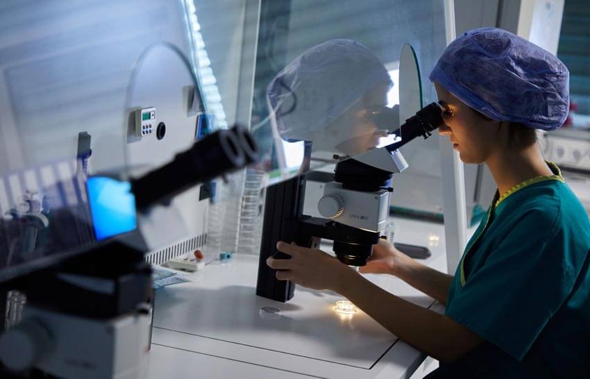
The sample can then be collected at the clinic and processed in the laboratory using sperm preparation or capacitation techniques, which aim to select a suitable population of sperm for fertilisation. The eggs retrieved from the donor are then fertilised using the appropriate technique, which may be conventional IVF or intracytoplasmic sperm injection (ICSI), depending on the circumstances.
If donor sperm is used instead, the arrangements are different: the sample comes from a sperm bank and is selected according to the relevant medical, legal and compatibility criteria. In both cases, the laboratory coordinates fertilisation and embryo culture while the recipient's endometrium is prepared to receive the embryo.
Discover the laboratory stages:
IVF treatment with donor eggs, step by step.
Generally, no. Endometrial preparation mainly involves hormonal medication, so the recipient does not need ovarian stimulation injections or an egg retrieval procedure, two of the most physically demanding stages of IVF using one's own eggs.
Oestrogen can be taken as tablets or patches, while progesterone is often administered vaginally. As with any hormonal medication, side effects may occur, including breast tenderness, bloating, abdominal discomfort, vaginal discharge when progesterone is used locally, or mild mood changes. Their severity varies between patients.
Embryo transfer is not usually painful either. Some women describe discomfort similar to that experienced during a gynaecological examination, particularly when the speculum is inserted, but anaesthesia or sedation is not normally required.
Yes, in most cases. Endometrial preparation does not require bed rest or time off work, unless a particular medical circumstance means otherwise. Patients can therefore walk, work, drive and maintain their usual routine in the weeks leading up to transfer. Moreover, as there is no ovarian stimulation, the ovaries do not become enlarged as they do during conventional IVF. This means the same restrictions relating to vigorous exercise or the risk of ovarian torsion associated with stimulation do not apply.
The most important things at this stage are to take medication at the prescribed times and attend scheduled monitoring appointments.
Madero S, Rodriguez A, Vassena R, Vernaeve V.
Endometrial preparation: effect of estrogen dose and administration route on reproductive outcomes in oocyte donation cycles with fresh embryo transfer.
.
2016;
1755-1764
Estudio sobre el efecto de la dosis y la vía de administración de estrógenos en los resultados reproductivos de ciclos de ovodonación con transferencia embrionaria en fresco:
https://pubmed.ncbi.nlm.nih.gov/27141040/
Younis JS, Simon A, Laufer N.
Endometrial preparation: lessons from oocyte donation.
.
1996;
873-884
Revisión científica sobre la preparación endometrial mediante estrógenos y progesterona en tratamientos de ovodonación y su influencia en la receptividad del endometrio:
https://pubmed.ncbi.nlm.nih.gov/8941049/
Arce H, Velilla E, López-Teijón M.
Association between endometrial thickness in oocyte donation cycles and pregnancy success rates.
.
2016;
1288-1294
Estudio que analiza la relación entre el grosor endometrial y las tasas de embarazo en ciclos de ovodonación, evaluando su utilidad como indicador del éxito reproductivo:
https://pubmed.ncbi.nlm.nih.gov/25707405/
Check JH, Nowroozi K, Choe J, Dietterich C.
Endometrial thickness is predictive of histologic endometrial maturation in women undergoing hormone replacement for ovum donation.
.
1996;
380-383
Investigación sobre la relación entre el grosor endometrial y la maduración histológica del endometrio en mujeres que reciben tratamiento hormonal para ovodonación:
https://pubmed.ncbi.nlm.nih.gov/8751733/
Remohí J, Gutiérrez A, Cano F, Ruiz A, Simón C, Pellicer A.
Long oestradiol replacement in an oocyte donation programme.
.
1995;
1387-1391
Estudio sobre los efectos de prolongar la administración de estradiol durante la preparación endometrial en receptoras de ovocitos y su relación con la implantación embrionaria:
https://pubmed.ncbi.nlm.nih.gov/7593502/
Borini A, Dal Prato L, Bianchi L, Violini F, Cattoli M, Flamigni C.
Effect of duration of estradiol replacement on the outcome of oocyte donation.
.
2001;
185-190
Investigación sobre la influencia de la duración del tratamiento con estradiol en la receptividad endometrial y los resultados de embarazo en ciclos de donación de ovocitos:
https://pubmed.ncbi.nlm.nih.gov/11432108/
Michalas S, Loutradis D, Drakakis P, Kallianidis K, Milingos S, Deligeoroglou E.
A flexible protocol for the induction of recipient endometrial cycles in an oocyte donation programme.
.
1996;
1063-1066
Estudio que evalúa un protocolo flexible de preparación endometrial con estrógenos y progesterona para coordinar la transferencia embrionaria en tratamientos de ovodonación:
https://pubmed.ncbi.nlm.nih.gov/8671391/
Gursu T, Goksever Celik H, Eraslan A, Angun B, Ozaltin S, Yeh J.
Impact of endometrial thickness change in response to progesterone on live birth rates embryo transfers with fresh oocyte donation cycles.
.
2022;
3260-3267
Estudio sobre los cambios en el grosor endometrial tras la administración de progesterona y su posible relación con las tasas de nacido vivo en ciclos de ovodonación:
https://pubmed.ncbi.nlm.nih.gov/35983690/
American Society for Reproductive Medicine (ASRM).
Gamete and embryo donation guidance.
.
2024;
799-813
Guía clínica de la ASRM que recoge recomendaciones sobre la selección y evaluación de donantes, los criterios médicos y genéticos y la seguridad de los procedimientos de donación de gametos y embriones:
https://www.asrm.org/practice-guidance/practice-committee-documents/guidance-regarding-gamete-and-embryo-donation/
Privacy Policy
IVF-LIFE, hereby informs you of its privacy policy applied to personal data. The use of the website does not imply that the user must provide any information regarding their identity, and cannot be associated with a specific user by the company. In the event that personal data is provided, this data will be collected and used in accordance with the limitations and rights set out in the aforementioned regulation.
WHO IS RESPONSIBLE FOR THE PROCESSING OF YOUR DATA?
DATA CONTROLLER: IVF SPAIN ALICANTE S.L.U (hereinafter IVF-Life)
CIF: B-54973227
Postal Address: Avenida de Ansaldo, no. 13, 03540, Alicante (Alicante)
Contact with the Data Protection Officer: dpo@ivf-life-group.com
DATA CONTROLLER: Instituto Vasco de Fertilidad (hereinafter IVF-Life)
CIF: B95575874
Postal Address: Avenida de Tolosa, 71-73, 20018 Donostia / San Sebastián (Spain)
Contact with the Data Protection Officer: dpo@ivf-life-group.com
DATA CONTROLLER: IVF Spain Madrid (hereinafter IVF-Life)
CIF: B 85138113
Postal Address: C/ de Manuel de Falla, 6-8, 28036 Madrid (Spain)
Contact with the Data Protection Officer: dpo@ivf-life-group.com
WHAT TYPE OF DATA DO WE PROCESS?
Depending on the products, services, or functionalities that you wish to enjoy, we may need to process different types of data. In general, this data will be, depending on the case, the following:
- Your identification data: for example: name, surname, language, country from which you interact with us, contact details, etc.
- Connection, geolocation and navigation data.
- Commercial information if you are subscribed to our newsletter.
- Data about your tastes and preferences.
WHAT IS THE ORIGIN OF THE DATA?
The use of certain services or requests addressed to IVF-LIFE are subject to the prior completion of the corresponding form. All the information provided by the User through the forms on the Portal for the above purposes or any other must be truthful.
To this end, the User guarantees the authenticity of all the data provided and will keep the information provided to IVF-LIFE perfectly up to date so that it corresponds at all times to the User's real situation. In any case, the User will be solely responsible for any false or inaccurate statements made and for any damage caused to IVF-LIFE or third parties because of the information provided.
By clicking on the "send" button on the corresponding form, the User expressly consents to his/her data being used to send professional communications about his/her query by e-mail or by any other means.
However, the User may revoke the consent given and oppose the sending of such communications in each of the communications received or by sending them to the address indicated in this legal notice and enabled for this purpose. Likewise, in each of the communications sent, the procedure for opposing the sending of new professional communications shall be expressly indicated.
Users who provide personal data of other natural persons are obliged to comply, in relation to said data, with all obligations deriving from the data protection regulations in force, and in particular the duty to inform and obtain the consent of the owner of the personal data.
When we request your personal data to provide access to certain website functionalities, we will mark the required fields to ensure the service can be delivered.
Please note that if you choose not to provide us with this data, you may not be able to complete your registration as a user or you may not be able to enjoy those services or functionalities.
For the processing of data, IVF-LIFE adopts the security levels required by current European and national legislation on the Protection of Personal Data. Notwithstanding the above, technical security in a medium such as the Internet is not impregnable and there may be leaks due to the malicious actions of third parties.
Our services are aimed at people of legal age, in the event that some of our services are specifically aimed at minors, IVF-LIFE will request the consent of parents or guardians for the collection of personal data or, where appropriate, for the automated processing of data as set out in current legislation. If the User is a minor, they are required to have the prior consent of their parents or guardians before proceeding to the inclusion of their personal data in the forms on the Website.
Initially, your personal data are provided directly by you, but in certain cases they may originate from centres, referring doctors and public health systems with which there is collaboration.
FOR WHAT PURPOSES DO WE PROCESS YOUR PERSONAL DATA?
Depending on how you interact with our website, we will process your personal data for the following purposes:
To deal with requests or requests that you make through the different means incorporated into our website.
We only process the personal data that is strictly necessary to manage or resolve your request or application.
For marketing purposes
To the extent that you subscribe to our newsletter, your personal data will be used for the periodic sending of company newsletters.
You should remember that you can unsubscribe from the newsletter, at any time and free of charge, by contacting us via the e-mail address of the data controller or by following the instructions that we will provide you with in each of our communications.
For the management of social networks
If you follow us or contact us through the different social networks in which we are present, we will process the data that you provide to us through these for the sole purpose of responding to your messages or comments and managing our contact list.
Usability and quality analysis to improve our services (cookies).
If you access our platform, we inform you that we will process your browsing data for analytical and statistical purposes, i.e. to understand how users interact with our platform and thus be able to make improvements to it. Likewise, we sometimes carry out quality actions and surveys aimed at finding out the degree of satisfaction of our customers and users and detecting those areas in which we can improve.
WHAT IS THE LEGAL BASIS FOR PROCESSING YOUR DATA?
The legal basis that allows us to process your personal data also depends on the purpose for which we process them, so we will find different legal bases under the following purposes:
Attention to requests for information.
The legal basis for the processing of your data is the informed consent to respond to queries raised through the website, forms and other means provided, which implies the prior reading and acceptance of these conditions by the interested party.
Marketing
The legitimate basis for processing your data for marketing purposes is the consent you provide.
Social networking
The basis of legitimacy is the informed consent to respond to queries raised through the social network, as well as the time when you follow us and want to be part of our contact list.
Usability and quality analysis (cookies)
User consent to the use of cookies.
HOW LONG WILL WE KEEP YOUR DATA?
Request for information. We will process your data for as long as it is necessary to deal with your request or request and thereafter 1 year.
Marketing
We will process your data until you unsubscribe or cancel your subscription to the newsletter.
Social media
We will process your data for as long as it is necessary to deal with your request and for as long as you are a follower of our profile.
WILL WE SHARE YOUR DATA WITH THIRD PARTIES?
Personal data will not be transferred or communicated to third parties, except: legal obligation, we obtain your express consent, or we understand that there is a legitimate interest on our part, which will be duly communicated to you.
However, we inform you that we are present in the following social networks: Facebook, LinkedIn, Instagram and YouTube, to keep our followers informed regarding the company's news and services.
You can access these social networks voluntarily and through the links available on the website itself or by accessing directly and on your own from your browser or installed app.
You should know that this website is not responsible for the use of the data that these social networks may make. However, when these data are generated in the social network and derive from an interaction with our website, we will not be held responsible for the use of such data.
In order to consent to our transfer from this website and the processing of your data, you should carefully read the privacy policy of the social network you wish to access:
Facebook: Facebook Privacy Policy
Instagram: Instagram Privacy Policy
LinkedIn: Linkedin Privacy Policy
YouTube: Youtube Privacy Policy
Spotify: Spotify Privacy Policy
In this way, before clicking on the logos of the SS. RR. present on our website, you can inform yourself and make a redirection with prior knowledge of the processing of your data and on a voluntary basis.
Other technological services:
- WhatsApp: WhatsApp Privacy Policy
- Companies of the "IVF LIFE Group" on the legal basis of consent or legitimate interest: Instituto Vasco de Fertilidad S.L.U., with NIF B95575874 and address at Avda Tolosa, 71-73 Donostia (Gipuzkoa), and IVF Spain Madrid S.L., with NIF B85138113 and address at Calle Manuel de Falla, 6-8, 28036 Madrid.
WHAT ARE YOUR RIGHTS WHEN YOU PROVIDE US WITH YOUR DATA?
We undertake to respect the confidentiality of your personal data and to guarantee you the exercise of your rights.
You may exercise them free of charge by sending us an e-mail to our address above, simply by providing proof of identity, the reason for your request and the right you wish to exercise.
If we consider it necessary in order to identify you, we may ask you for a copy of a document proving your identity. In particular, regardless of the purpose or the legal basis on which we process your data, you have the right to:
Ask us for access to the data we hold about you.
Ask us to rectify the data we already hold about you. Please note that by actively providing us with your personal information in any way, you warrant that it is true and accurate and you undertake to notify us of any changes or modifications to it.
Ask us to delete your data to the extent that they are no longer necessary for the purpose for which we need to process them as we have previously informed you, or to the extent that we no longer have the legitimacy to do so.
Ask us to restrict the processing of your data, which means that in certain cases you may ask us to temporarily stop processing the data or to keep it longer than necessary when you may need it. If you have given us your consent to process your data for any purpose, you also have the right to withdraw your consent at any time.
To receive the personal data concerning you that you have provided to us in a structured, commonly used and machine-readable form and to transfer it to another data controller.
Moreover, where the processing of your data is based on our legitimate interest, you also have the right to object to the processing of your data.
Finally, we inform you of your right to lodge a complaint with the relevant data protection supervisory authority, with the Spanish Data Protection Agency.
CHANGES TO THE PRIVACY POLICY
We may change the information contained in this Privacy Policy from time to time. If we do so, we will notify you by various means through the Platform, or we will notify you at your email address when the change in question is significant to your privacy, so that you can review the changes, assess them and, where appropriate, object to or unsubscribe from any service or functionality.
INFORMATION ABOUT COOKIES
We use cookies and similar devices to facilitate your navigation on the Platform, to know how you interact with us and, in certain cases, to be able to show you advertising based on your browsing habits. Please read our Cookie Policy to learn more about the cookies and similar devices we use, their purpose and other information of interest.